
Knee Arthroscopy
Keyhole Surgery for Lasting Joint Health
Relieve painful joint locking, swelling, and tears with minimally invasive knee arthroscopy. Mr. Lebur Rohman specializes in advanced meniscal repair and targeted resection—restoring smooth, pain-free movement through precision keyhole care..
Knee Arthroscopy & Meniscal Surgery Guide
Minimally Invasive Keyhole Joint Diagnosis, Joint Preservation, Meniscal Repair, and Specialist Reconstruction under Mr. Lebur Rohman
1. Knee Arthroscopy: Precision Keyhole Surgery
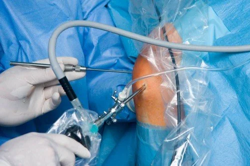
Knee Arthroscopy is a state-of-the-art, minimally invasive keyhole procedure used to inspect, diagnose, and treat mechanical conditions inside the knee joint. Through two tiny incisions (portals)—each measuring only 5 to 10 millimeters—a pencil-sized fiber-optic camera (arthroscope) and specialized micro-instruments are introduced into the joint. High-definition video is projected onto a surgical monitor, allowing Mr. Lebur Rohman to treat internal joint structures with absolute anatomical precision.
Why Perform Knee Arthroscopy?
Keyhole surgery provides unmatched diagnostic clarity and immediate surgical intervention for mechanical knee pain that fails to respond to non-surgical treatment.
Pathologies Treated
- Meniscal Tears & Displacements
- Loose Bone or Cartilage Fragments
- Articular Cartilage Flaps / Lesions
- Synovial Inflammation (Synovitis)
- Plica Syndrome & Stiffness
Key Patient Benefits
- Day-Case Procedure (Home Same Day)
- Minimal Soft Tissue Disruption
- Significantly Lower Post-Op Pain
- Lower Infection Risk (<0.1%)
- Accelerated Recovery Timeline
Experiencing Catching, Locking, or Persistent Knee Pain?
Don't let joint dysfunction hold you back. Consult with lower limb specialist Mr. Lebur Rohman for an accurate diagnosis and personalized preservation plan.
Book Your Consultation Today2. The Shift to Meniscus Preservation & Clinical Evidence
For decades, a torn meniscus was viewed as expendable and routinely removed. Contemporary orthopedic science recognizes this as a critical error. The meniscus is essential for load distribution, shock absorption, and protecting articular cartilage. Preserving meniscal tissue whenever possible is the single most impactful surgical decision for delaying or preventing long-term knee osteoarthritis.
3. Meniscal Vascularity & Evidence-Based Likelihood of Healing
The meniscus is divided into three distinct vascular zones. Because tissue healing relies entirely on blood supply, a tear's location dictates whether it can be successfully sutured or needs to be trimmed.
Red-Red Zone (Outer 10-30%)
Rich Blood Supply: High healing capacity (~85-95% success). Ideal for all-inside, inside-out, or outside-in suture repair.
Red-White Zone (Middle Third)
Intermediate Blood Supply: Moderate healing potential. Repair is prioritized in active patients, sometimes aided by biologic stimulation.
White-White Zone (Inner Third)
Avascular Region: Receives nutrients solely via synovial fluid. Tears here heal unreliably and are managed via partial meniscectomy.
4. Clinical Tear Patterns & Pathology Types
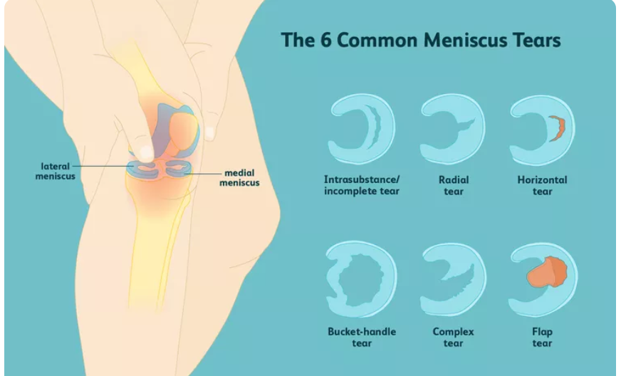
Meniscal tears are categorized by their physical geometry, depth, and cause (traumatic twisting versus age-related degeneration):
Common Tear Configurations
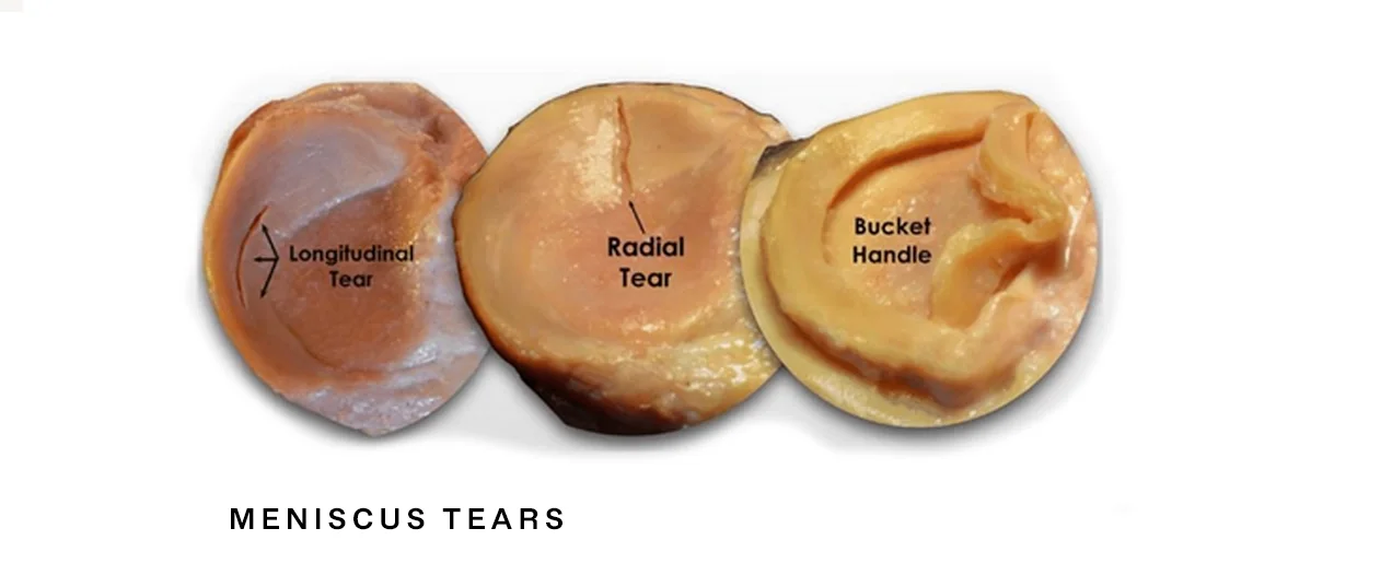
- Longitudinal / Vertical: Runs parallel to circumferential fibers; prime candidate for repair in vascular zones.
- Bucket-Handle Tear: A large longitudinal tear that flips into the joint notch, causing physical knee locking.
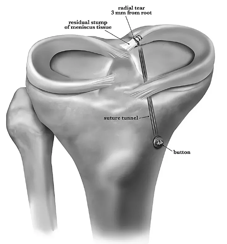
- Radial Tears: Cuts across circumferential fibers, disrupting the load-bearing hoop stress mechanism.
- Horizontal Cleavage: Splits cartilage into upper and lower layers; frequently associated with meniscal cysts.
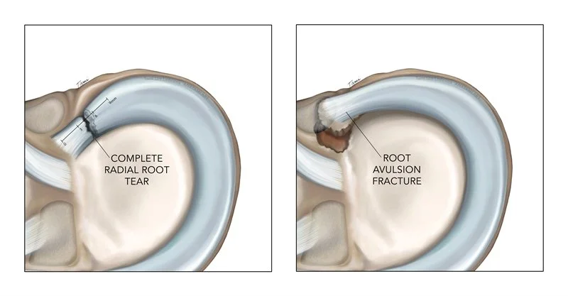
- Root Tears: Detachment at the bony root insertion; behaves functionally like losing the entire meniscus.
Degenerative & Non-Surgical Pathways
Degenerative Fraying: Common in mature knees. When mechanical locking is absent, conservative treatment is prioritized:
- Rest & Activity Modification: Avoiding aggravating pivot movements.
- Anti-Inflammatory Care: Targeted NSAIDs, ice packing, and compression.
- Physiotherapy: Strengthening quadriceps and hamstrings for stability.
- Intra-Articular Injections: Cortisone, Hyaluronic Acid, or PRP for symptomatic relief.
- Unloader Braces: Offloading stress from the affected compartment.
5. The Three Surgical Repair Techniques
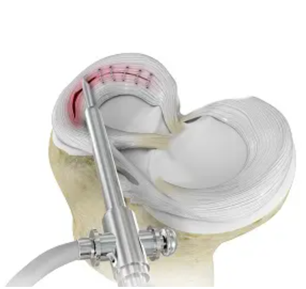
Keyhole meniscus repair matches specific surgical stitching techniques to the anatomical location and complexity of the tear:
| Technique | How It Works | Best Tear Location | Reported Success Rate | Invasiveness & Profile |
|---|---|---|---|---|
| All-Inside | Sutures & anchors placed entirely within joint via arthroscopy. | Posterior horn / posterior third | ~80% – 93% | Least invasive; no extra incision needed. |
| Inside-Out | Sutures passed inside-to-outside, tied over capsule via small side incision. | Mid-body & large bucket-handle tears | ~80% – 100% (Gold Standard) | Requires small accessory safety incision. |
| Outside-In | Sutures passed outside-to-inside, tied externally over capsule. | Anterior horn / anterior third | ~86% | Excellent control for front-of-knee tears. |
6. Post-Meniscectomy Care & Meniscal Transplantation (MAT)
For young or active patients who have previously undergone a complete or subtotal meniscectomy and now experience persistent compartment pain without advanced osteoarthritis, Meniscal Allograft Transplantation (MAT) offers a biological solution. MAT replaces the missing meniscus with a donor tissue graft to restore shock absorption and delay joint breakdown.
*Note: Meniscal Allograft Transplantation is a specialized biological reconstruction. Explore our dedicated Meniscal Transplant section for full details on eligibility, donor matching, and recovery protocols.*
7. Frequently Asked Questions Regarding Knee Arthroscopy
How should I prepare for knee arthroscopy keyhole surgery?
What should I expect on the day of my day-case knee arthroscopy procedure?
What is the recovery timeline following a Partial Meniscectomy (meniscus trimming)?
- Weight-Bearing: You can place full weight on your leg immediately, using crutches for 2 to 5 days for comfort.
- Driving: Usually permitted after 1 to 2 weeks once you can perform an emergency stop safely.
- Work & Activity: Desk work can resume within a few days; physical work and sports usually resume at 3 to 4 weeks.
What is the recovery timeline and brace protocol following a Meniscal Repair?
- Protective Brace: You will wear a hinged knee brace locked between 0-90 degrees for 6 weeks to prevent deep bending.
- Weight-Bearing Restrictions: Full weight-bearing with crutches is often fine from day 1. Deep squats beyond 90 degrees are restricted for up to 3 months.
- Full Recovery: Return to high-impact sports or heavy manual work typically takes 4 to 6 months. Patients can start to drive at 6 weeks once the brace is removed.
What initial Range of Motion (ROM) exercises should I perform after surgery?
- Ankle Pumps: Actively flex and extend your foot up and down to encourage venous blood circulation and prevent DVT.
- Static Quadriceps Sets: Lie flat, tighten your thigh muscle, push the back of your knee firmly into the bed, hold for 5 seconds, and release.
- Seated Heel Slides: Sit on a firm chair, slide your foot back gently under the chair to bend the knee comfortably, hold for 5 seconds, and straighten.
- Straight Leg Raises: Tighten your thigh, lift your leg 6 inches off the bed, hold for 3 seconds, and slowly lower.
Where can I access guided physiotherapy booklets and video resources for meniscal recovery?
Access official rehabilitation guides and video demonstrations below:
Take the First Step Toward Pain-Free Mobility
Get an expert assessment from Consultant Orthopaedic Surgeon Mr. Lebur Rohman to preserve your joint health and return to active living.
Schedule Your Consultation