Cartilage Surgery
Advanced Cellular Cartilage Repair.
Repair cartilage defects before arthritis takes hold. Discover how cutting-edge biological techniques (AutoCart, AMIC & ACI) regenerate your knee’s natural surface without joint replacement.
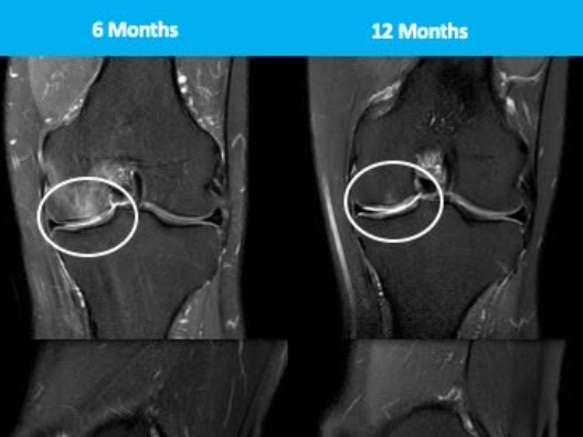
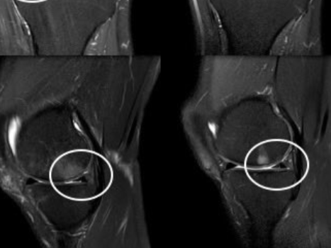
Advanced Biological Cartilage Surgery
Regenerative Knee Preservation & Defect Repair under Specialist Knee Surgeon Mr. Lebur Rohman
National & Regional Lead in Cartilage Restoration
- National Specialist Expertise: Mr. Rohman is one of approximately 22 recognized cartilage surgeons nationwide performing complex biological joint restoration.
- Regional Expertise: One of only 2 specialist cartilage surgeons in the North East of England.
- Regional Leadership: Co-Lead of the upcoming North East Regional Cartilage Centre — Gateshead Cartilage Centre.
1. Understanding Knee Cartilage Damage (Pathophysiology)
Why Cartilage Cannot Heal on Its Own
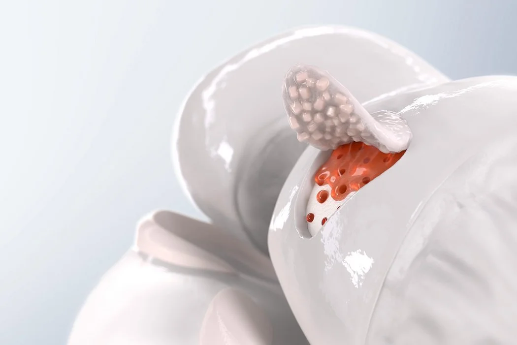
Articular cartilage is the ultra-smooth, slippery white tissue that coats the ends of your bones in the knee joint. It acts as a friction-free cushion, allowing bones to glide smoothly against each other during movement.
Unlike skin, muscle, or bone, articular cartilage is avascular—meaning it has no direct blood supply. It also lacks nerves and lymphatic drainage. When a pothole or defect is created in the cartilage (due to a sports injury, impact, or twisting force), the body cannot deliver stem cells, oxygen, or growth factors through the bloodstream to repair it. Over time, an untreated pothole exposes the underlying bone, leading to friction, inflammation, catching, and eventual early-onset osteoarthritis.
2. Advanced Cartilage Regeneration Procedures
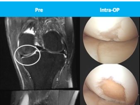
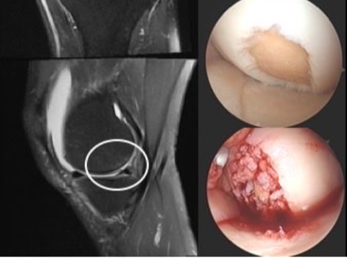
Biological cartilage repair focuses on filling the "pothole" with new, healthy tissue to restore a smooth joint surface and stop progressive joint breakdown.
1. AutoCart (Minced Cartilage Implantation)
What it is: A modern, single-stage biological repair procedure.
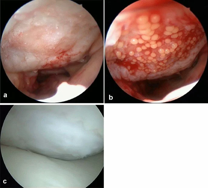
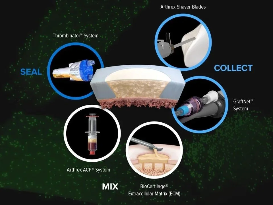
How it works: Small pieces of healthy, non-weight-bearing cartilage are harvested from the patient's own knee, "minced" into tiny micro-fragments, and mixed with Autologous Thrombin or Platelet-Rich Plasma (PRP) derived from the patient’s blood. This creates a natural biological putty that is glued precisely into the cartilage defect.
Why it works: Mincing the cartilage breaks open the dense tissue matrix, releasing living cartilage cells (chondrocytes) that migrate, multiply, and synthesize new, smooth cartilage within the defect.
2. AMIC (Autologous Matrix-Induced Chondrogenesis)
What it is: A single-stage technique combining microfracture with a natural collagen scaffold.
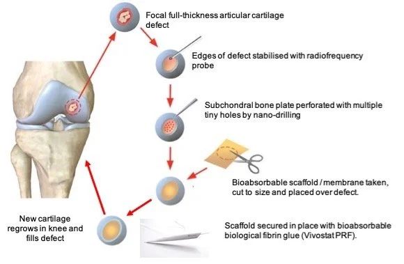
How it works: Tiny micro-punctures are made in the bone base at the bottom of the cartilage defect to release stem cells and blood from the bone marrow. A specialized collagen matrix/membrane is cut to shape and secured over the defect to cover and trap the stem cells in place.
Why it works: The collagen membrane acts as a protective "blanket" that prevents the stem cells from washing away, guiding them to differentiate into durable repair tissue.
3. ACI (Autologous Chondrocyte Implantation)
What it is: A traditional, two-stage cell therapy procedure.
How it works:
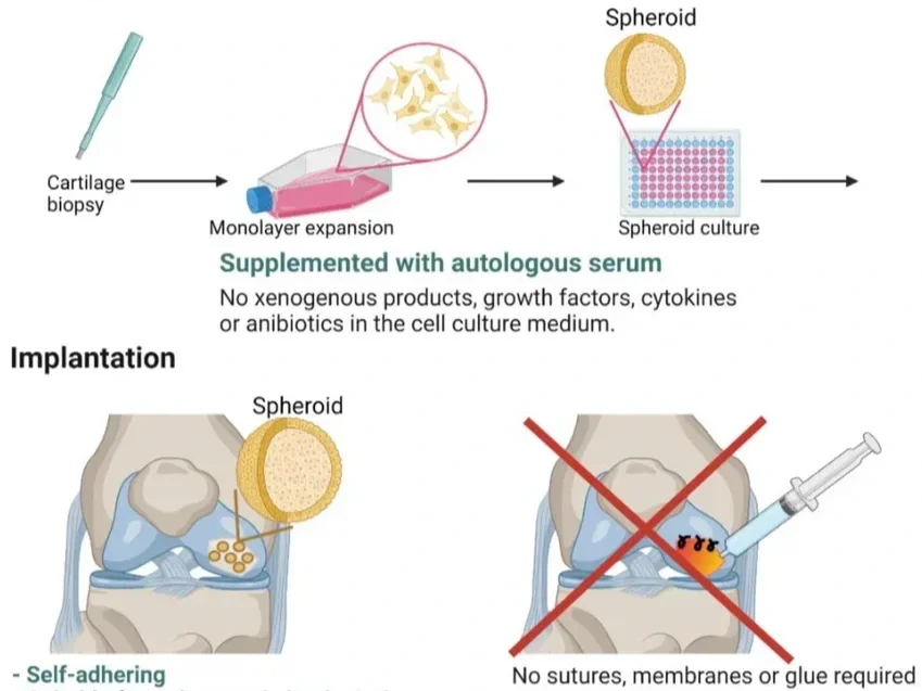
Stage 1: A keyhole procedure harvests a tiny biopsy of healthy cartilage, which is sent to a specialized laboratory where the cartilage cells are cultured and grown into millions of cells over several weeks.
Stage 2: A second operation implants the cultured cells under a patch or membrane secured over the defect.
Why it works: Provides a massive concentration of pure, laboratory-expanded living cartilage cells directly into large defects.
Suffering from Isolated Knee Cartilage Damage?
Get evaluated by national cartilage specialist Mr. Lebur Rohman to explore biological joint restoration.
Book Specialist Assessment →3. Evidence-Based Comparison & Pros/Cons
Each biological procedure offers distinct advantages depending on the defect size, location, patient age, and surgical history:
| Procedure | Surgical Stages | Clinical Indications | Pros (+) | Cons (-) |
|---|---|---|---|---|
| AutoCart (Minced Cartilage) | Single-Stage (1 Operation) | Small to medium focal defects (1–4 cm²). Excellent for active patients wanting a single intervention. |
|
|
| AMIC (Matrix-Induced Chondrogenesis) | Single-Stage (1 Operation) | Small to moderate defect areas (1.5–5 cm²). Ideal when bone marrow stimulation is desired alongside scaffold stabilization. |
|
|
| ACI / MACI (Chondrocyte Implantation) | Two-Stage (2 Operations) | Large or complex focal defects (>4–8 cm²), or failing previous cartilage repair procedures. |
|
|
4. Frequently Asked Questions & Sample Rehabilitation Protocols
What is Autologous Chondrocyte Implantation (ACI / MACI) and how does the two-stage process work?
Autologous Chondrocyte Implantation (ACI) is a state-of-the-art cell therapy procedure designed for larger or complex cartilage defects (>4 cm²). It requires two separate surgical stages:
- Stage 1 (Biopsy Harvest): A minor keyhole arthroscopy is performed to assess the joint and harvest a tiny, rice-grain-sized piece of healthy, non-weight-bearing cartilage. This sample is sent under sterile conditions to a licensed cell laboratory, where the chondrocytes (cartilage cells) are extracted and grown into millions of living cells over 3 to 6 weeks.
- Stage 2 (Cell Implantation): Once the cell culture is ready, a open or mini-open procedure is performed. The defect is cleaned, and the laboratory-expanded chondrocytes—either suspended or seeded onto a 3D collagen membrane (MACI)—are implanted into the defect and glued securely with fibrin sealant.
How does ACI compare to single-stage procedures like AutoCart and AMIC?
The primary difference lies in the number of surgeries, cell concentration, and defect size capability:
- ACI: Requires 2 surgeries and laboratory cell expansion. It provides the highest concentration of pure cartilage cells and is the clinical gold standard for very large defects (>4–8 cm²).
- AutoCart: A single-stage surgery using minced native cartilage fragments mixed with the patient's own blood products (PRP/thrombin). It avoids a second operation and laboratory fees, making it ideal for small-to-medium defects.
- AMIC: A single-stage surgery utilizing bone marrow stem cells released via microfracture, stabilized under a 3D collagen patch. It is best suited for small-to-moderate defects where marrow stimulation is appropriate.
What is the sample rehabilitation protocol for AutoCart (Minced Cartilage)?
AutoCart rehabilitation balances protecting the soft biological matrix while encouraging chondrocyte matrix production through early motion:
AutoCart Sample Rehabilitation Protocol:
- Weeks 0–2: Touch weight-bearing (10–15 kg) with crutches. Knee brace locked in extension for walking. Passive Range of Motion (PROM) 0°–60° daily using Continuous Passive Motion (CPM) or manual therapy.
- Weeks 3–6: Partial weight-bearing (50%), progressing to full weight-bearing by Week 6 as tolerated. Brace unlocked (0°–90°). Begin stationary cycling with zero resistance.
- Weeks 7–12: Wean off brace and crutches. Full joint range of motion. Progressive quadriceps and closed-chain strengthening exercises.
- Months 3–6: Low-impact cardio (swimming, rowing, outdoor cycling). Dynamic stability and proprioception training.
- Months 6–9+: Return-to-running assessment and gradual resumption of non-contact pivoting sports.
What is the sample rehabilitation protocol for AMIC surgery?
AMIC rehabilitation focuses on shielding the stem cell blood clot and collagen patch from shear stress:
AMIC Sample Rehabilitation Protocol:
- Weeks 0–6: Strict partial or touch weight-bearing with crutches (depending on femoral vs. patellar location). Protective hinged brace worn at all times. Controlled passive flexion restricted to 0°–90° for the first 4 weeks to prevent patch displacement.
- Weeks 6–12: Gradual transition to 100% weight-bearing. Wean off brace. Focus on active knee extension, patellar mobility, and gluteal/quadriceps activation.
- Months 3–6: Resistance training, leg press (0°–60°), elliptical trainer, and hydrotherapy.
- Months 9–12: High-level functional agility drills, plyometrics, and assessment for return to impact activities.
What is the sample rehabilitation protocol for ACI / MACI surgery?
ACI requires the longest and most structured recovery because cultured cells undergo distinct biological phases (tissue synthesis, maturation, and remodeling):
ACI / MACI Sample Rehabilitation Protocol:
- Weeks 0–6 (Protection Phase): Touch weight-bearing (10–15% body weight) with crutches. CPM machine used 4–6 hours daily (0°–90°) to nourish cultured cells without mechanical load.
- Weeks 6–12 (Transition Phase): Gradual weight-bearing progression (25% increase per week). Wean off crutches by Week 10–12. Hydrotherapy and stationary bike with light resistance.
- Months 3–6 (Remodeling Phase): Full weight-bearing, closed-kinetic-chain exercises (squats 0°–60°), gait retraining, and core/pelvic stability.
- Months 6–12 (Maturation Phase): Advanced strengthening, outdoor cycling, straight-line jogging starting around Month 8–9 upon MRI confirmation of defect fill.
- Months 12–18: Full return to competitive pivoting sports and high-impact manual labor.
What are the risks and long-term success rates of biological cartilage repair?
Success Rates: Long-term clinical trials confirm significant pain reduction, restored joint smooth-gliding, and functional recovery in 80% to 85% of properly selected patients, delaying joint replacement for over a decade.
Risks: Infection (<1%), deep vein thrombosis (DVT), graft hypertrophy (overgrowth of repair tissue), joint stiffness, or graft non-healing/delamination (~10–15%).
Preserve Your Joint with National Subspecialist Care
Schedule a private evaluation with Mr. Lebur Rohman to discuss biological AutoCart, AMIC, or ACI cartilage restoration.
Book Private Assessment →